Looking for the best way to ace your Life Sciences grade 12 exam? This comprehensive revision guide combines Life Sciences grade 12 study notes, Life Sciences grade 12 Past Exam questions, and topic summaries to help you prepare for tests and exams with confidence. Boost your marks by accessing Life Sciences grade 12 free downloadable exam preparation resources, including Life Sciences grade 12 , study guides, and detailed solutions.
The human nervous system
The human nervous system is a highly organised network that allows us to sense, process, and respond to the world around us. It is divided into two main parts: the central nervous system (CNS) and the peripheral nervous system (PNS). Together, they ensure that the body can receive information, interpret it, and react appropriately to keep us alive, safe, and functioning efficiently.
The Central Nervous System
The central nervous system (CNS) forms the body’s primary control centre, responsible for processing information, making decisions, and coordinating actions. It is made up of two main parts: the brain and the spinal cord. Together, these structures receive sensory input from the rest of the body, interpret it, and send out instructions to help us move, think, and react appropriately to the environment. To protect these delicate and vital parts, the brain is securely enclosed within the skull, and the spinal cord lies inside the vertebral column (backbone). Both are further wrapped in three protective membranes called the meninges, which consist of the tough outer dura mater, the web-like arachnoid layer, and the delicate pia mater closest to the nervous tissue. These layers, along with the cerebrospinal fluid they help contain, cushion the CNS against shock, reduce friction, and protect it from infection and physical injury.
The Brain
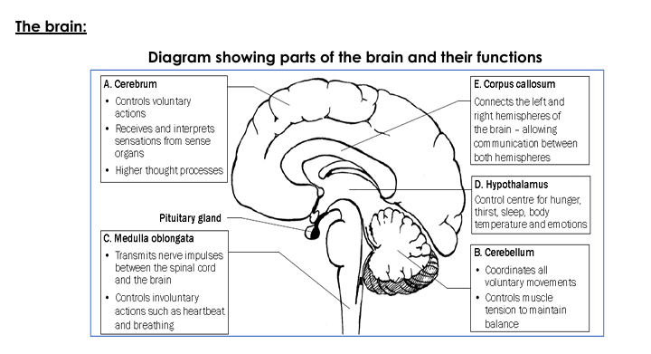
The brain is the largest and most complex organ of the central nervous system. It acts as the control centre for thought, memory, emotion, and voluntary and involuntary activities. One major part of the brain is the cerebrum, which is the biggest section and handles higher mental functions such as thinking, reasoning, language, and memory. The cerebrum is divided into two large parts called hemispheres, which are linked by a thick bundle of nerve fibres known as the corpus callosum. This connection ensures that the left and right hemispheres can share information and coordinate activities effectively.
The cerebrum is the largest and most complex part of the human brain, responsible for higher mental processes and voluntary activities. It allows us to think, reason, plan, speak, write, and remember, and it processes sensory input so that we can interpret what we see, hear, feel, taste, and smell. Its outer layer, the cerebral cortex, is divided into specialised lobes: the frontal lobe handles decision-making and movement control; the parietal lobe manages touch and spatial awareness; the temporal lobe is involved in hearing and memory; and the occipital lobe focuses on vision. Together, these areas enable us to understand and interact with the world around us in a thoughtful and coordinated way.
Connecting the two hemispheres of the cerebrum is the corpus callosum, a thick, C-shaped band of nerve fibres. This structure acts as a communication bridge between the left and right sides of the brain, making it possible to share information quickly. Thanks to the corpus callosum, both hemispheres can work together smoothly — for example, allowing us to combine logical thinking with creativity or coordinate movements that involve both sides of the body.
Beneath the cerebrum lies the cerebellum, which is essential for coordinating voluntary muscle movements. It ensures our movements are smooth and balanced, and it plays a crucial role in helping us maintain posture and equilibrium — for instance, keeping us steady when we walk on uneven ground or when we quickly change direction.
Near the centre of the brain is the thalamus, which serves as a vital relay station for sensory and motor signals. It receives information coming from the eyes, ears, skin, and other parts of the body, then directs these messages to the right areas of the cerebral cortex for interpretation. This process helps us become consciously aware of our surroundings and react appropriately.
Just below the thalamus is the hypothalamus, a small but powerful control centre. The hypothalamus maintains the body’s internal balance (homeostasis) by regulating body temperature, thirst, hunger, and daily biological rhythms. Importantly, it also connects the nervous system to the endocrine system by telling the pituitary gland when to release hormones, affecting growth, metabolism, and stress responses.
Attached to the base of the brain is the pituitary gland, often called the “master gland” because it produces hormones that influence many other glands in the body. Through its hormone release, it helps control growth, development, metabolism, and reproductive processes, making it key to healthy body function and development.
The pineal gland, a small, pea-shaped structure deeper in the brain, plays a role in regulating our sleep–wake cycle. It does this by producing the hormone melatonin, which rises and falls in response to light and darkness, helping to set our natural body clock or circadian rhythm.
Forming part of the brainstem is the pons, which lies above the medulla oblongata and below the midbrain. The pons helps relay signals between the cerebrum and cerebellum and plays a role in controlling breathing. It also carries sensory information up to the thalamus, contributing to our awareness and coordination.
At the base of the brain, connecting to the spinal cord, is the medulla oblongata. This part controls critical involuntary reflexes, such as regulating the heartbeat, controlling breathing rhythm, and managing reflex actions like swallowing and sneezing. Because it keeps these vital functions running automatically, the medulla oblongata is essential for life.
Looking for the best way to ace your Life Sciences grade 12 exam? This comprehensive revision guide combines Life Sciences grade 12 study notes, Life Sciences grade 12 Past Exam questions, and topic summaries to help you prepare for tests and exams with confidence. Boost your marks by accessing Life Sciences grade 12 free downloadable exam preparation resources, including Life Sciences grade 12 , study guides, and detailed solutions.
The Spinal Cord
The spinal cord is a long, cylindrical structure that forms the second major part of the central nervous system (CNS), alongside the brain. It begins at the medulla oblongata, which is located at the base of the brain, and travels downward through the vertebral canal formed by the stacked vertebrae of the spine, until it reaches the lumbar region of the back. This protective bony covering helps shield the delicate nerve tissues inside.
The spinal cord’s primary role is to act as the main communication pathway between the brain and the rest of the body. It carries sensory information from receptors (such as those detecting pain, temperature, and touch) up to the brain and delivers motor commands from the brain down to the muscles and glands, enabling voluntary movements and essential body functions.
In addition, the spinal cord handles reflex actions, which are fast, automatic responses that do not first pass through the brain, like quickly pulling your hand away from something hot. This combination of relaying information and reflex control helps the body respond quickly and remain balanced in changing conditions.
Structurally, the spinal cord is organised in a way that supports its vital functions:
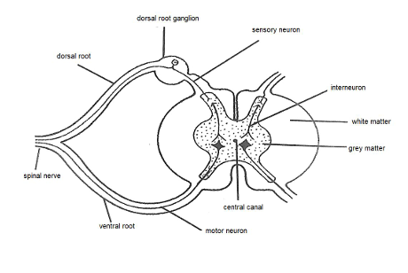
At the centre of the spinal cord is a narrow central canal, which runs the entire length of the cord and is filled with cerebrospinal fluid (CSF). This fluid acts as a natural cushion to protect the spinal cord from sudden shocks, helps distribute nutrients, remove waste products, and maintains the chemical balance and pressure needed for nerve cells to work properly.
The spinal cord also contains two main types of tissue: grey matter and white matter. The grey matter forms the butterfly-shaped inner core and is mainly made of neuron cell bodies, dendrites, and supporting glial cells. It is especially important for processing information and coordinating reflex actions. Surrounding it is the white matter, which is made up of myelinated nerve fibres (axons). The myelin covering makes signals travel faster, allowing quick communication between the brain and the body.
Along its length, the spinal cord gives rise to spinal nerves, which extend outward to connect the CNS with muscles, skin, and organs throughout the body. Each spinal nerve arises from both sides of the spinal cord and is formed by the merging of two distinct roots:
The dorsal root, which contains sensory neurons. These neurons carry incoming information from the body into the spinal cord, helping us detect sensations such as heat, pain, and pressure.
The ventral root, which consists of motor neurons. These neurons transmit outgoing signals from the spinal cord to muscles and glands, producing movement and other bodily responses.
The Peripheral Nervous System (PNS)
The peripheral nervous system (PNS) is a major division of the human nervous system that includes all nervous tissue located outside the central nervous system (CNS), which itself consists of the brain and spinal cord. The PNS acts as a vast communication network that connects the CNS to the rest of the body, making sure that sensory information from the environment and internal body conditions reach the brain and spinal cord, and that motor instructions from the CNS reach muscles and glands. Without the PNS, the CNS would be isolated and unable to control or receive feedback from the body’s organs, limbs, and tissues.
The peripheral nervous system is made up of two main sets of nerves: 12 pairs of cranial nerves that emerge directly from the brain, and 31 pairs of spinal nerves that branch out from the spinal cord to reach almost every part of the body.
The cranial nerves play especially important roles in the head and neck areas, helping with senses like smell, sight, hearing, and taste, as well as movements like facial expression and eye movement.
The spinal nerves, in turn, serve areas of the torso and limbs, allowing us to move, feel sensations, and respond to changes in our environment.
Sensory and Motor Nerves
Within the peripheral nervous system (PNS), there are two main types of nerves that help maintain communication between the central nervous system (CNS) and the rest of the body: sensory nerves and motor nerves.
Sensory nerves play a vital role by carrying electrical signals from sensory receptors located in the skin, muscles, joints and internal organs towards the CNS. These signals allow us to experience different sensations, including touch, pain, temperature and pressure. For example, when you accidentally touch a sharp object, sensory nerves quickly transmit this information to the brain, making you aware of the pain.
Motor nerves, on the other hand, have the opposite role: they carry instructions from the CNS to the muscles and glands throughout the body. Thanks to motor nerves, we are able to perform both small and large movements, such as writing with a pen, walking, or even speaking. Together, sensory and motor nerves create a two-way communication network that ensures the body can sense, react and adapt to the environment effectively.
Looking for the best way to ace your Life Sciences grade 12 exam? This comprehensive revision guide combines Life Sciences grade 12 study notes, Life Sciences grade 12 Past Exam questions, and topic summaries to help you prepare for tests and exams with confidence. Boost your marks by accessing Life Sciences grade 12 free downloadable exam preparation resources, including Life Sciences grade 12 , study guides, and detailed solutions.
Subdivisions of the Peripheral Nervous System
The PNS is further divided into two functional subdivisions, each responsible for managing different types of body activities:
The somatic nervous system (SNS)
The autonomic nervous system (ANS)
This division helps the body coordinate both voluntary actions that we consciously control and involuntary actions that happen automatically.
Somatic Nervous System
The somatic nervous system is specifically designed to control voluntary movements of the skeletal muscles. It carries nerve impulses from the CNS to the muscles we can choose to move, allowing us to perform tasks like running, dancing, writing or picking up objects. For instance, when you decide to stand up and walk across a room, it is the somatic nervous system that transmits these instructions from your brain to your leg muscles, making the movement possible. This system gives us conscious control over our actions and allows us to respond purposefully to our environment.
Autonomic Nervous System
In contrast, the autonomic nervous system manages activities in the body that occur without conscious thought. It conducts nerve impulses from the CNS to involuntary muscles (such as the muscles lining the stomach and intestines, or the muscles in blood vessels) and to glands. This system ensures that essential processes — including heart rate, digestion, breathing rhythm, blinking of the eyes and reflex actions like sneezing — happen smoothly and automatically.
Functions of the peripheral nervous system:
Conducting Impulses from Receptors to the Central Nervous System
One of the main roles of the peripheral nervous system (PNS) is to carry impulses from sensory receptors to the central nervous system (CNS). These receptors are located all over the body, including in the skin, muscles, joints, and internal organs. They detect changes both inside and outside the body, such as temperature, touch, pain, and pressure. By transmitting these sensory signals to the CNS, the PNS keeps the brain and spinal cord constantly updated about the environment and the body’s internal state. This function is vital because it helps us sense what is happening around us, supports reflex actions, and allows the CNS to process information and decide on suitable responses for survival and daily living.
Conducting Impulses from the Central Nervous System to Effectors
Another key function of the peripheral nervous system is to carry impulses from the central nervous system to effectors, which are muscles and glands in the body. After the CNS processes sensory information and decides on an appropriate reaction, it sends motor commands out through the PNS. These commands reach skeletal muscles, allowing us to perform voluntary movements like walking, writing, or lifting objects. At the same time, they can stimulate glands to produce substances such as saliva, sweat, or hormones needed for various processes. This pathway ensures that decisions made by the brain and spinal cord translate into coordinated physical actions and responses, enabling us to interact with our environment, maintain balance, and keep bodily functions working properly.
Location and Functions of the Autonomic Nervous System
The autonomic nervous system (ANS) is a branch of the peripheral nervous system (PNS). Unlike the somatic nervous system, which controls voluntary actions, the ANS manages involuntary actions—those that happen automatically without conscious control. It consists of nerve pathways that extend from the central nervous system (the brain and spinal cord) to smooth muscles, cardiac muscles, and glands throughout the body. Because of this, the ANS plays a critical role in regulating functions like heart rate, breathing, digestion, and pupil size, ensuring the body stays balanced and responsive to changing internal and external conditions.
Subdivisions of the Autonomic Nervous System
The autonomic nervous system has two main subdivisions:
The sympathetic division
The parasympathetic division
These divisions often have opposite effects on target organs, which helps the body maintain homeostasis by balancing activity levels according to different situations.
Sympathetic Division: Preparing the Body for Emergencies
The sympathetic division becomes most active when the body faces stress, danger, or any situation requiring quick action—often described as the “fight or flight” response. When activated, it prepares the body by:
Increasing the heart rate, so more blood and oxygen reach muscles
Raising blood pressure to ensure rapid circulation
Dilating (widening) the pupils to let in more light and improve vision These changes make the body alert, focused, and ready to respond to emergencies, whether that means escaping danger or facing a physical challenge.
Parasympathetic Division: Returning the Body to Normal
The parasympathetic division helps the body relax and recover once the emergency or stressful situation has passed. This division supports “rest and digest” activities by:
Slowing down the heart rate back to normal
Lowering blood pressure
Constricting (narrowing) the pupils This helps conserve energy, promote digestion, and maintain daily bodily functions essential for health and recovery.
Looking for the best way to ace your Life Sciences grade 12 exam? This comprehensive revision guide combines Life Sciences grade 12 study notes, Life Sciences grade 12 Past Exam questions, and topic summaries to help you prepare for tests and exams with confidence. Boost your marks by accessing Life Sciences grade 12 free downloadable exam preparation resources, including Life Sciences grade 12 , study guides, and Life Sciences grade 12 detailed solutions.