This content is tested repeatedly in Grade 12 examinations, both directly and indirectly, in questions on DNA, inheritance, protein synthesis, and genetics. Learners who master this work are able to answer definition, short factual, diagram-based, and application questions accurately and confidently.
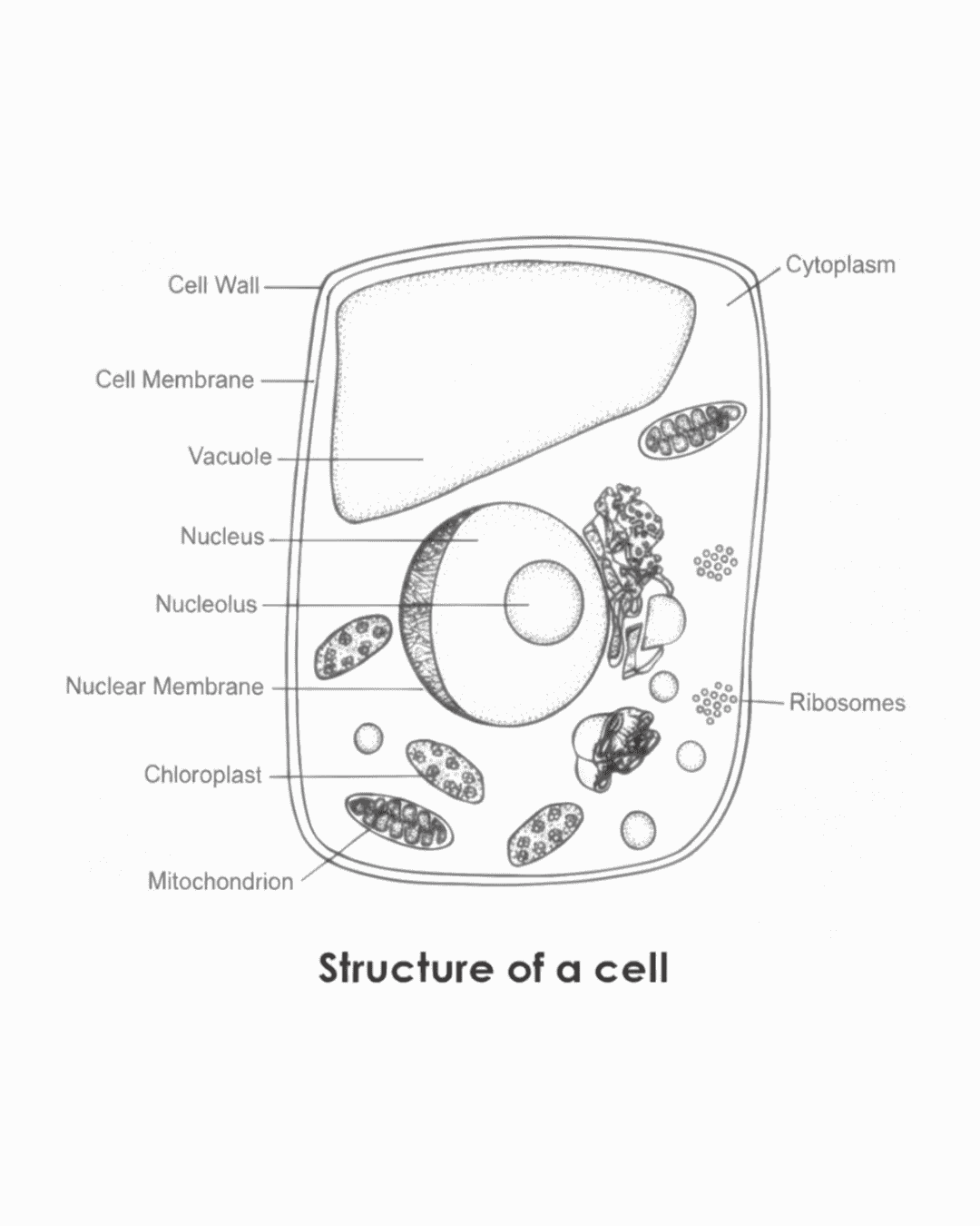
Revision of the structure of the cell
Emphasis on cytoplasm, ribosomes and the nucleus
Cytoplasm
The cytoplasm is the semi-fluid substance found inside the cell membrane in which organelles are suspended and where most metabolic reactions of the cell take place.
The cytoplasm consists partly of a liquid portion and partly of a jelly-like portion.
Functions of the cytoplasm
Stores substances required by the cell
Allows metabolic reactions to take place
Learners lose marks if they omit metabolic reactions or confuse the cytoplasm with the nucleus.
Ribosomes
Ribosomes are small, non-membranous structures composed of RNA and proteins that occur in both plant and animal cells.
Location of ribosomes
Free-floating in the cytoplasm
Attached to the endoplasmic reticulum
Function of ribosomes Ribosomes are the sites of protein synthesis.
Learners lose marks if they state that ribosomes produce energy or contain DNA.
Structure of the nucleus
The nucleus is a large membrane-bound organelle that controls the activities of the cell and contains the genetic material.
Parts of the nucleus
A double nuclear membrane
Nuclear pores
Nucleoplasm
A nucleolus
A chromatin network
The chromatin network shortens and thickens to form chromosomes during cell division.
Functions of the nucleus
Controls the activities of the cell
Contains chromosomes that carry hereditary characteristics
Learners lose marks if they fail to link chromatin to chromosomes or confuse the nucleolus with the nucleus.
Nucleic acids
Nucleic acids are large biological molecules that store and transmit genetic information in cells.
Types of nucleic acids
DNA (deoxyribonucleic acid)
RNA (ribonucleic acid)
DNA carries the genetic information of an organism and determines inherited characteristics.
RNA is involved in protein synthesis and carries genetic information from DNA to the ribosomes.
Learners must not confuse the roles of DNA and RNA.
Nucleotides
Nucleic acids consist of repeating subunits called nucleotides.
Structure of a nucleotide
A nitrogenous base
A sugar molecule
A phosphate group
Learners lose marks if any component is omitted.
Location of DNA
DNA is mainly located in the nucleus, where it forms part of the chromatin network.
Chromosomes are thread-like structures found in the nucleus and are composed of DNA wrapped around proteins called histones.
A gene is a short segment of a DNA molecule that codes for a specific inherited characteristic.
DNA outside the nucleus
In the mitochondria of plant and animal cells
In the chloroplasts of plant cells
Learners lose marks if they state that DNA occurs only in the nucleus.
Full-mark model answers
Name the two types of nucleic acids. DNA and RNA.
State what nucleic acids consist of. Nucleic acids consist of nucleotides.
State where DNA is found in a eukaryotic cell. DNA is found mainly in the nucleus and in smaller amounts in mitochondria and chloroplasts.
Define a gene. A gene is a short segment of a DNA molecule that codes for a specific inherited characteristic.
Distinction checkpoint
A learner who can read, understand, and accurately reproduce the content above will be able to score full marks on all Term 1 Week 1 questions related to cell structure, nucleic acids, and the location of DNA.
Brief history of the discovery of DNA
Exam relevance
This topic is commonly assessed in short factual questions, sequencing questions, and source-based questions. Examiners reward correct names, correct dates, correct contributions, and correct sequencing. Any inaccuracy leads to mark loss.
Key scientists and contributions
Rosalind Franklin and Maurice Wilkins (1952)
Rosalind Franklin and her assistant Maurice Wilkins conducted research on the structure of DNA using X-ray diffraction techniques.
The X-ray diffraction images provided evidence that:
DNA has a regular, repeating structure
DNA is arranged in a helical form
Learners lose marks if they state that Franklin discovered the structure of DNA or if they omit the X-ray diffraction method.
Watson and Crick (independent research)
James Watson and Francis Crick conducted independent research on DNA.
They used:
Existing experimental data
X-ray diffraction evidence
to develop a model of DNA structure.
Learners lose marks if they state that Watson and Crick carried out X-ray diffraction themselves.
Proposal of the DNA model (1953)
In 1953, Watson and Crick proposed a three-dimensional double helix model for the structure of DNA.
The model explained:
The shape of the DNA molecule
How genetic information could be stored
Learners lose marks if they omit the term double helix or the year 1953.
Nobel Prize recognition (1962)
In 1962:
Watson and Crick received the Nobel Prize for the discovery of the structure of DNA
Maurice Wilkins also received the award for his contribution through X-ray photography
Learners lose marks if they include Rosalind Franklin as a Nobel Prize recipient or give the incorrect year.
Chronological summary
1952 – Rosalind Franklin and Maurice Wilkins produced X-ray diffraction images of DNA. 1953 – Watson and Crick proposed the three-dimensional double helix model of DNA. 1962 – Watson, Crick, and Wilkins received the Nobel Prize for the discovery of the structure of DNA.
Full-mark model answers
Name the technique used by Franklin and Wilkins to investigate DNA. X-ray diffraction.
State the contribution of Watson and Crick to the discovery of DNA. They proposed a three-dimensional double helix model for the structure of DNA.
In which year was the structure of DNA proposed? 1953.
Why did Rosalind Franklin not receive a Nobel Prize? She had passed away before the Nobel Prize was awarded.
Common learner errors and mark loss
Learners lose marks if they:
Confuse who proposed the DNA model
Attribute the Nobel Prize incorrectly
Omit years
Use vague wording such as “helped discover DNA”
Precision is required.
Distinction checkpoint
A learner who can accurately reproduce the names, dates, methods, and sequence above will score full marks on all NSC questions related to the discovery of DNA.
Structure of DNA
DNA is a polymer. This means that DNA is a large molecule made up of many repeating smaller units joined together in a chain. In examinations, learners must be able to use the term polymer correctly, as it links directly to the idea that DNA is made of repeating units.
Key exam point DNA is described as a polymer because it consists of many repeating subunits.
DNA consists of two strands. Each strand is a long chain made up of repeating units. The two strands run alongside each other and together form one DNA molecule.
Common exam check Learners must state that DNA is double-stranded, not single-stranded.
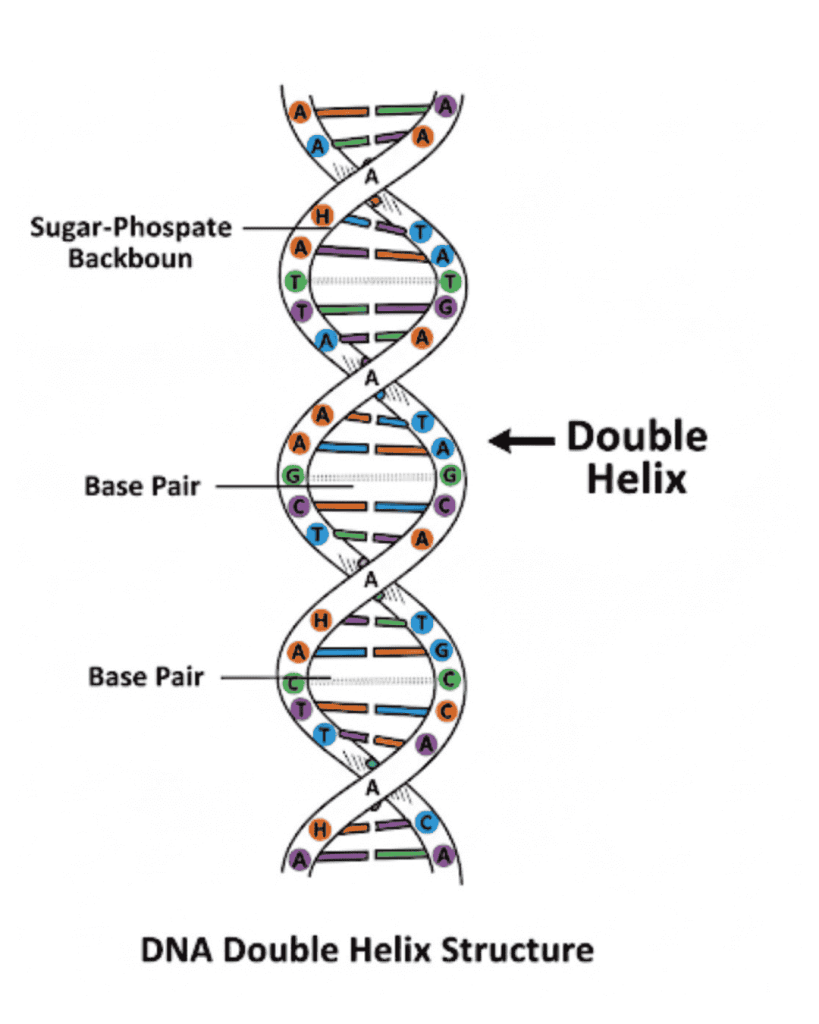
The two strands are twisted around each other. This twisting gives DNA its natural shape, known as a double helix. The term double helix must be used exactly in examinations, as it is the accepted scientific description of the shape of DNA.
Definition to memorise The natural shape of the DNA molecule is a double helix.
Diagram A labelled diagram of the DNA double helix
Two strands twisted around each other
A ladder-like appearance
Distinction checkpoint A learner should be able to write the following statements accurately in an exam:
The monomers of DNA are known as nucleotides. This means that nucleotides are the small repeating units that join together to form the DNA molecule. In examinations, learners must be able to correctly link the term monomer to nucleotide.
Key exam point DNA is a polymer made up of repeating monomers called nucleotides.
Each DNA strand consists of a long sequence of nucleotides joined together. The order of these nucleotides is important because it determines the genetic information carried by the DNA molecule.
Examiner focus Learners often lose marks by stating that nucleotides are made of amino acids. This is incorrect.
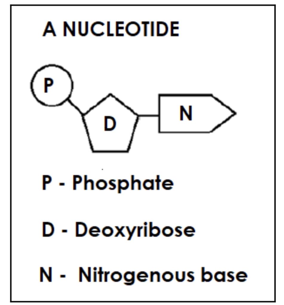
Structure of a nucleotide
Each nucleotide consists of three components. All three must be stated correctly to earn full marks.
A sugar molecule called deoxyribose
A phosphate group
A nitrogenous base
Definition to memorise A nucleotide consists of a deoxyribose sugar, a phosphate group, and a nitrogenous base.
The sugar and phosphate form part of the backbone of the DNA molecule, while the nitrogenous bases project inward and pair with each other.
Diagram Diagram of a single DNA nucleotide here.
This diagram is commonly used in examinations to test identification and labelling skills.
Distinction checkpoint A distinction-level learner must be able to write:
The monomers of DNA are nucleotides.
Each nucleotide consists of a sugar, a phosphate group, and a nitrogenous base.
Structure of DNA: detailed organisation of the molecule
DNA is made up of repeating units called nucleotides. Each nucleotide is organised in a specific way that gives DNA its unique structure and function.
Structure of a nucleotide
Each nucleotide consists of three components:
A deoxyribose sugar
A phosphate group
One nitrogenous base
The nitrogenous base combines with the deoxyribose sugar, while the deoxyribose sugar combines with the phosphate group. These linkages are essential for forming the structure of the DNA molecule.
The sugar and phosphate do not attach randomly. They combine in a fixed, repeating pattern that forms the framework of DNA.
Sugar–phosphate backbone of DNA
The sides of the DNA ladder consist of alternating deoxyribose sugar molecules and phosphate groups.
This repeating sugar–phosphate pattern forms the backbone of the DNA molecule and provides strength and stability to the structure.
When describing DNA structure in an examination, learners must refer to the sides as alternating deoxyribose and phosphate groups.
Nitrogenous bases in DNA
There are four types of nitrogenous bases found in DNA:
Adenine (A)
Guanine (G)
Cytosine (C)
Thymine (T)
Each nucleotide contains only one of these nitrogenous bases.
Complementary base pairing
Nitrogenous bases pair in a specific and fixed manner, known as complementary base pairing:
Adenine always combines with Thymine
Guanine always combines with Cytosine
As a result:
There are equal numbers of adenine and thymine bases
There are equal numbers of guanine and cytosine bases
This relationship is written as:
A = T and G = C
Because of this pairing, one DNA strand is the complement of the other strand.
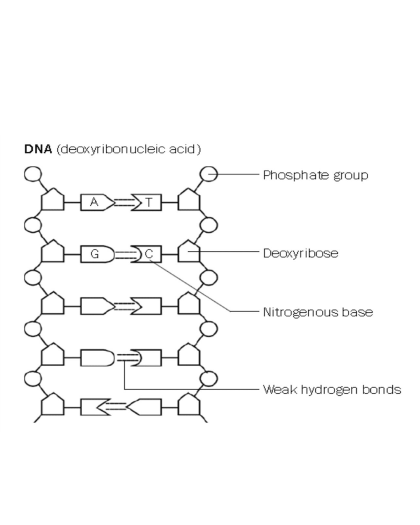
Hydrogen bonds between bases
The paired nitrogenous bases are held together by weak hydrogen bonds.
These hydrogen bonds:
Hold the two DNA strands together
Are easily broken by enzyme action, which is essential during processes such as DNA replication
Learners lose marks if they describe these bonds as strong or fail to mention enzyme action.
Diagram of DNA structure
Alternating deoxyribose and phosphate groups forming the sides
Nitrogenous bases forming the rungs
Adenine paired with thymine
Guanine paired with cytosine
Hydrogen bonds between paired bases
Distinction checkpoint
A learner who can accurately reproduce the structural relationships, base-pairing rules, and bonding described above will be able to score full marks on all DNA structure questions.
How to recognise a DNA molecule
A DNA molecule can be identified by the following structural features: • Double-stranded molecule • Contains the nitrogenous base thymine (T) instead of uracil (U) • A always joins with T • G always joins with C
Functions of DNA
DNA performs two essential functions in living cells. These functions explain why DNA is fundamental to inheritance and cell activity, and they are commonly tested in short factual and explanation questions.
DNA and hereditary information
Sections of the DNA molecule form genes.
A gene is a section of DNA that carries information for a specific inherited characteristic.
These genes:
Carry hereditary information
Are passed from parents to offspring
Learners lose marks if they describe genes as whole chromosomes or confuse genes with traits.
DNA and protein synthesis
DNA contains coded information for protein synthesis.
This means that DNA stores instructions that determine:
Which proteins are made
The structure and function of those proteins
Proteins are essential because they control cell structure and cell activities.
Learners lose marks if they state that DNA makes proteins directly.
DNA replication
DNA replication is a fundamental cellular process that ensures genetic information is passed accurately from one cell to another.
Definition of DNA replication
DNA replication is the process during which a DNA molecule makes an exact copy (replica) of itself.
This definition must be memorised exactly, as it is frequently assessed in definition questions.
When DNA replication takes place
DNA replication takes place during interphase of the cell cycle.
This ensures that:
Each new cell receives a complete and identical copy of DNA
Learners lose marks if they state that DNA replication occurs during mitosis.
Diagram: DNA Replication
Screenshot
Process of DNA replication
DNA replication occurs in a specific sequence of steps. These steps must be written in the correct order to earn full marks.
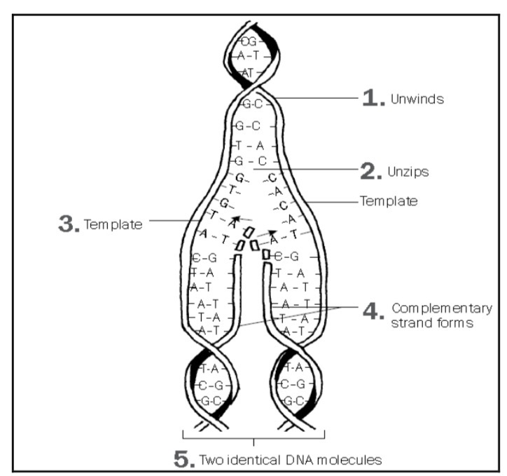
Step 1: Unwinding of the DNA molecule
The DNA double helix unwinds.
This exposes the nitrogenous bases.
Step 2: Breaking of hydrogen bonds
The weak hydrogen bonds between the nitrogenous bases break.
As a result, the two DNA strands separate and unzip.
Learners lose marks if they describe strong bonds breaking.
Step 3: Template strands
Each of the original DNA strands serves as a template.
This means each strand is used as a pattern to build a new complementary strand.
Step 4: Formation of complementary strands
Free nucleotides present in the nucleoplasm attach to the exposed bases on each template strand.
Base pairing occurs as follows:
Adenine pairs with Thymine
Guanine pairs with Cytosine
This ensures accurate copying of genetic information.
Step 5: Formation of identical DNA molecules
Two identical DNA molecules are formed.
Each DNA molecule consists of:
One original (parent) strand
One newly formed strand
This is known as semi-conservative replication.
Learners lose marks if they state that both strands are newly formed.
Diagram of DNA replication
Diagram interpretation questions are common in examinations.
Distinction checkpoint
A learner who can:
State the functions of DNA
Define DNA replication accurately
Describe the process of DNA replication in the correct sequence
Explain how identical DNA molecules are formed
will be able to score full marks on DNA replication questions.
Significance of DNA replication
DNA replication is essential for growth, repair, and reproduction in living organisms. Its significance explains why DNA must be copied accurately before cell division.
Doubling of genetic material
DNA replication doubles the genetic material in a cell.
This ensures that:
Each new cell receives the same amount of DNA
Genetic information is maintained during cell division
Learners lose marks if they state that DNA replication halves genetic material.
Formation of identical daughter cells
DNA replication results in the formation of identical daughter cells during mitosis.
This is important because:
All daughter cells have the same genetic information
Cell functions remain consistent after division
Learners lose marks if they confuse mitosis with meiosis in this context.
DNA profiling
DNA profiling is a technique used to compare DNA samples and identify individuals based on their genetic material.
Uniqueness of DNA profiles
Every person, except identical twins, has a unique DNA profile.
This uniqueness allows DNA to be used reliably for identification purposes.
Description of a DNA profile
A DNA profile can be described as an arrangement of black bars.
These black bars:
Represent DNA fragments of a person
Form a pattern that is specific to that individual
Learners lose marks if they describe a DNA profile as the DNA sequence itself.
Uses of DNA profiling
DNA profiling can be used for several important purposes:
As proof of paternity
To trace missing persons
To identify genetic disorders
To establish family relations
To match tissues for organ transplants
To identify dead persons or animals
To identify crime suspects in forensic investigations
Correct use of terminology is required when listing applications.
Common errors made by learners in examinations
Learners often lose marks because they are:
Unable to answer and interpret questions based on the structure of DNA, DNA replication, and DNA profiling
Unable to make appropriate drawings and label diagrams correctly
Practising diagram drawing and interpretation is essential for examination success.
Distinction checkpoint
A learner who can explain the significance of DNA replication, describe DNA profiling accurately, list its uses correctly, and avoid common examination errors will be able to score full marks on questions related to these topics.